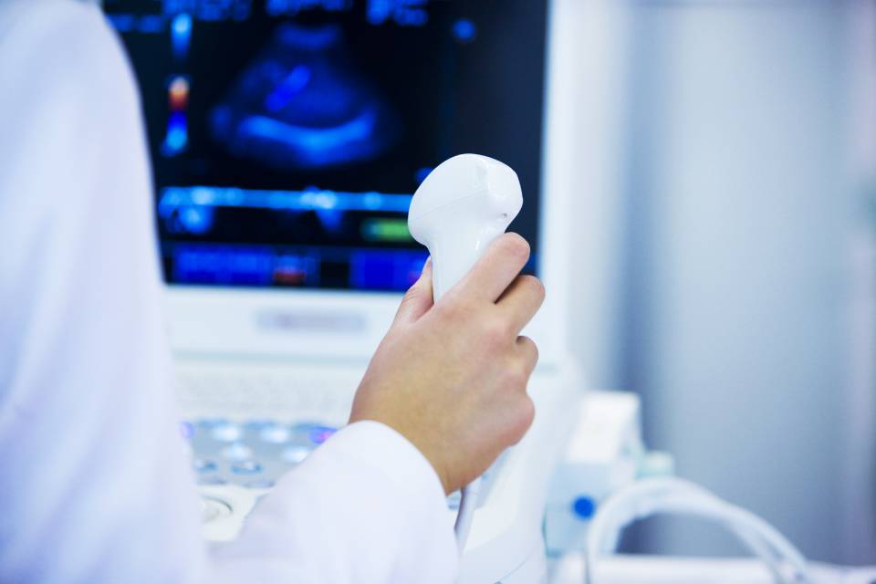
Ultrasound and color Doppler ultrasound

Ultrasound and Color Doppler in Cosenza

Among the various diagnostic services offered by the Federico Radiology practice in Cosenza we also find ultrasound examinations and color Doppler ultrasound, a non-invasive examination used to further analyze any pathologies of veins and arteries.

Contact us

Ultrasound

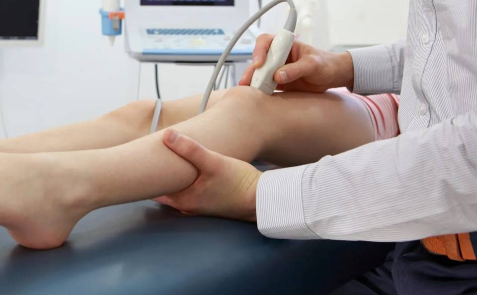
Ultrasound scans are based on ultrasound and the transmission of ultrasound waves.For this exam, booking is mandatory, even if waiting times are short.
We perform:
  • Upper abdominal ultrasound

    examine the liver, gallbladder, biliary tract, spleen, kidneys, abdominal aorta. For a correct execution of the test it is advisable to fast for at least 6 hours.

  • Lower abdomen ultrasound

    it is possible to examine the structure of the bladder, prostate, uterus and appendages. For this exam it is recommended to drink 1 liter of water half an hour before the booking time.

  • Complete abdominal ultrasound

    both the upper and lower abdomen are examined. You must have fasted for 6 hours and you must drink 1 liter of water half an hour before your appointment.

  • Transrectal prostate ultrasound

    through an internal probe it is possible to visualize the prostate directly and obtain information on the size and characteristics of the gland. The evening before you must prepare for the exam, which will be communicated at the time of booking.

  • Transvaginal ultrasound

    it is a diagnostic imaging technique that investigates the morphology and state of health of the female internal genital organs. Thanks to this test it is possible to study the uterus, ovaries and appendages directly.

  • Breast ultrasound

    studies the gland and breast tissue and any pathological alterations, both benign such as cysts, fibroadenomas, and malignant such as tumors.

  • Axillary cable ultrasound

    breast cancer tumor cells can often migrate along the lymphatic pathways and therefore localize in the lymph nodes; the first lymph node stations involved are almost always located at the level of the axillary cable.

  • Muscle tendon ultrasound

    investigates muscle and tendon pathologies, highlighting any tears, contractures, distractions, tendonitis, cysts, bursitis, subcutaneous or intramuscular hematomas of the elbow, knee, foot, hand, shoulder, wrist, ankle, Achilles tendon.

  • Thyroid and neck ultrasound

    specifically, the diameters of both lobes of the thyroid and its shape are measured, the presence of nodules and their characteristics are verified. The ultrasound study of the neck also allows the lymph node stations located in the neck area to be studied in particular the supraclavicular, lateralcervical, submandibular ones and the salivary glands, which have an important function in the immune system.

  • Ultrasound of superficial lymph nodes

    studies the lymphatic nodules present in the neck, armpits and groin. It allows us to highlight any lymphadenopathies and evaluate their reactive or malignant nature.

  • Ultrasound of the skin and subcutaneous tissue

    it must be performed when you want to analyze a visible or palpable neoformation to be better defined and referred to cysts, lipomas, inflammatory changes, thickness of subcutaneous fat

  • Ultrasound of the testicles

    allows the volumetric and morphological evaluation of the site of the testicles, epididymis and provides information on the structure of the testicles (for example the presence of tumors) and on any pathologies of the testicular appendages (epididymis cysts, hydrocele etc..)

  • Pediatric ultrasound of the hips

    It is a very important test for early diagnosis of a congenital developmental anomaly of the newborn's hip. It is advisable to do this test from the fourth/fifth week of life and within the third month.

  • Neonatal gastroesophageal ultrasound

    Reflux is a very common disorder in children, especially in the first months of life. It is the ascent into the esophagus of acidic material coming from the stomach. In newborns it manifests itself with regurgitation and emission of saliva, mucus and milk from the mouth. Before starting the exam the child must eat a full milk meal. The number of episodes of gastro-esophageal reflux is counted by evaluating the extent and considering the duration of stagnation in the esophagus. In newborns, observation is extended to the pyloric valve, located between the stomach and duodenum, to study hypertrophic pyloric stenosis. The latter requires surgical treatment.

Ecocolordoppler

EcocolorDoppler is a latest generation tool for ultrasound diagnostics. Absolutely non-invasive, it allows ultrasound visualization of the main blood vessels and the study of the blood flow within them.
  • Eco-color Doppler renale

    it is a valid diagnostic tool in the study of hypertension and unknown renal dysfunction.

  • Echo-color Doppler of the abdominal aorta and great vessels

    It is carried out to analyze any aneurysmal pathologies. Highlights wall thickening, thrombus or evidence of dissection. The test is performed after a diet period of at least three days; this reduces the presence of air and liquids in the intestine, facilitating the passage of ultrasound. Usually the right and left renal artery, the aorta under the diaphragm and the iliac district are explored. Vascular pathologies such as arterial and venous stenosis, aneurysms, thrombosis and venous insufficiency are studied. The test serves to reveal any aneurysms (dilations) or stenosis and occlusions (narrowings) of the large abdominal blood vessels. Pathologies that often develop without providing particular symptoms. This is why it is important to undergo this test at age 60, even if you are well.

  • Eco-color-Doppler tiroideo

    it serves to show the morphology of the gland and determine its volume by checking the presence of nodules and blood flow.

  • Echo-color-Doppler spermatic vessels

    also called scrotal or testicular doppler, it is a non-invasive diagnostic test that allows you to evaluate the venous and arterial vascularization of the testicles. In particular, it is performed to diagnose varicocele.

  • Arterial and venous echo-color Doppler of the lower limbs

    this exam allows you to have a complete picture of the venous and arterial system of the legs and serves to evaluate the circulatory system. Typically this test is prescribed to verify the presence of obstructions or emboli and their actual location; for any thrombosis or to determine the widening of an artery, also identify the source and severity of varicose veins or detect any tumors or congenital malformations.

  • Arterial and venous echo-color Doppler of the upper limbs

    the venous and arterial circulatory system of the arms as well as the legs is examined.

  • Echo-color-Doppler of the supra-aortic trunks (TSA)

    : through this test it is possible to obtain images relating to the large arteries that arise from the aorta and carry blood to the upper limbs, head and brain (carotid and vertebral arteries). This test diagnoses arterial diseases. Among these pathologies, atherosclerosis, characterized by the presence of obstructing plaques, develops very slowly and can remain silent for many years, until the size of the plaques significantly hinders the passage of blood or, due to the fragility of the structure of the plaques themselves, the detachment of emboli may occur which can block smaller arteries; it is in these cases that stroke or cerebral ischemia occurs.

Contact us to book an ultrasound or color Doppler ultrasound
Contacts
Share by: